Goblet Cells, Cilia and Airway Defence
FREE BIOLOGY REVISION · UNIT 1 · A3.1.2
Pearson BTEC Level 3 AAQ Applied Science · Issue 7, May 2026
Revision notes
Coordinated protection
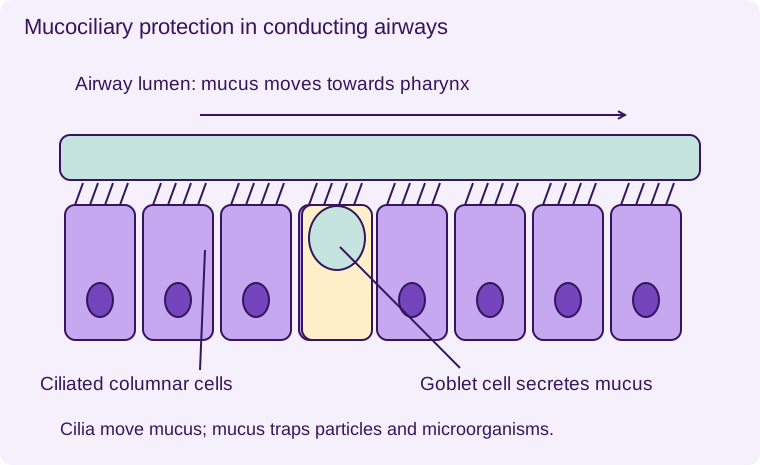
Much of the conducting respiratory tract is lined by ciliated columnar epithelium, often pseudostratified in larger airways. Columnar cells are taller than they are wide. Goblet cells secrete mucus that traps inhaled particles and microorganisms. Ciliated cells have many motile cilia on their exposed surface. Coordinated beating moves the mucus towards the pharynx, where it can be swallowed or expelled.
This mucociliary clearance reduces the number of pathogens reaching deeper lung surfaces. Mucus traps; cilia move. The two cell types have complementary roles, and neither should be described as directly producing antibodies. Swallowed material can be exposed to stomach acid, but trapping a microorganism does not instantly kill it.
Cellular machinery
Goblet cells synthesise and package the protein-containing components of mucus using ribosomes, RER, Golgi and secretory vesicles. Exocytosis releases their product. Ciliated cells contain mitochondria to supply ATP for movement. Cilia are microtubule-based structures extending from the cell surface; they are not root hairs or microvilli. Microvilli mainly increase surface area and do not produce the same coordinated sweeping movement.
Disruption and experimental reasoning
Smoke and other irritants can damage cilia and increase mucus production. When clearance falls, mucus and trapped organisms remain longer, increasing obstruction and infection risk. Coughing can partly compensate but does not restore a healthy epithelial surface.
Compare a healthy and damaged airway using mucus movement, ciliary coverage and infection frequency. In a controlled model, measure how far marked particles move in a known time. Control temperature and fluid viscosity because both affect movement. Use repeated samples rather than assuming one patch of tissue represents the entire airway. Do not infer that all respiratory surfaces contain goblet cells: the alveolar barrier must remain thin for diffusion.
- 1
- Mucus layer
- 2
- Cilia
- 3
- Goblet cell secreting mucus
- 4
- Ciliated columnar epithelial cells
Cilia move mucus towards the pharynx. Mucus traps particles and microorganisms. Schematic, not to scale.
Worked application and data skills
Marked particles travel 6 mm in 30 s over healthy model tissue and 2 mm in 30 s over damaged tissue. Speeds are 0.20 mm s⁻¹ and 0.067 mm s⁻¹. The damaged sample has about one-third the clearance speed. A plausible mechanism is reduced ciliary activity, but further measurements are needed to distinguish ciliary loss from changes in mucus viscosity.
Common misconceptions
Goblet cells secrete mucus; cilia move it. Microvilli and cilia have different functions. Alveoli do not use a thick mucociliary lining.
Examination tips
Name both cell types and explain their linked roles in preventing pathogens reaching gas-exchange surfaces.
Check your understanding
Five original questions. Use Tab to move between controls, arrow keys within a radio group, and Enter or Space on buttons. Feedback appears after Check Answer. Try Again clears the question. Attempts are not saved.
Sources and scope
Scope: Unit 1 A3.1.2. Original BTECRevision notes, schematics and questions. Independent resource; not endorsed by Pearson.
Official Pearson specification. Scientific review references are listed in the accompanying QA report.
EXAM RESOURCES
Biology exam resources
Apply your knowledge with written practice.
Browse exam resources