The Fluid Mosaic Model of Cell Membranes
FREE BIOLOGY REVISION · UNIT 1 · C1.1
Pearson BTEC Level 3 AAQ Applied Science · Issue 7, May 2026
Revision notes
A dynamic bilayer
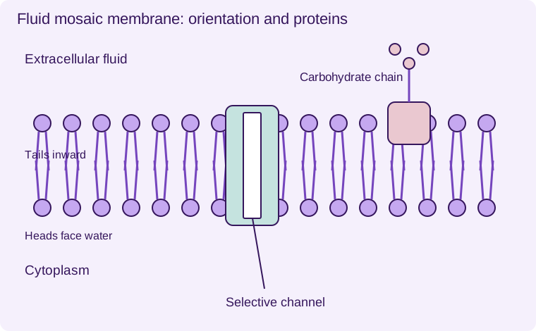
The cell-surface membrane consists principally of a phospholipid bilayer containing proteins and other components. Hydrophilic heads face the aqueous exterior and cytoplasm; hydrophobic tails face inward. This arrangement creates a barrier to many charged or strongly polar substances. Small non-polar molecules such as oxygen can cross the lipid region more readily.
Fluid describes lateral movement of many lipids and proteins. Mosaic describes the mixture and distribution of components. It does not mean every protein moves freely at the same speed: some are attached to cytoskeletal or extracellular structures. The membrane is neither a rigid wall nor a pair of protein sheets coating a lipid sandwich.
Proteins and other components
Channel proteins provide hydrophilic pathways for selected ions or water. Carrier proteins bind solutes and change conformation. Some carriers mediate facilitated diffusion; pumps support active transport. Receptor proteins detect signals and initiate responses. Enzymes associated with membranes catalyse reactions.
Glycoproteins and glycolipids have carbohydrate chains exposed on the extracellular face. They support recognition, adhesion and signalling. In animal membranes, cholesterol is interspersed among phospholipids and modulates fluidity and permeability; its effects depend on temperature. It is inaccurate to say that cholesterol simply makes every membrane rigid under all conditions.
Selective permeability
The bilayer’s hydrophobic core limits direct passage of ions and many polar solutes. Selective proteins allow controlled exchange. A membrane can therefore maintain different concentrations on either side. A cell wall provides support but does not substitute for this selective membrane.
Interpret evidence
Movement of labelled membrane components supports fluidity. Freeze-fracture studies reveal particles embedded in membranes, supporting the presence of proteins within the bilayer. Original schematics simplify molecular sizes and spacing; they are models rather than literal photographs. When annotating a diagram, show protein channels spanning the bilayer and carbohydrate chains on the correct face.
Changes in temperature, lipid composition or solvent exposure can alter permeability. A dye-leakage experiment must control tissue size, exposure time and washing to distinguish treatment effects from damaged cut edges.
- 1
- Hydrophilic heads face water
- 2
- Hydrophobic tails face inward
- 3
- Selective channel protein
- 4
- Extracellular carbohydrate chain
A schematic fluid mosaic membrane showing selected components; not all proteins or lipids are shown.
Worked application and data skills
A charged ion crosses poorly through a protein-free lipid bilayer but crosses rapidly when a selective channel is present and open. This supports a hydrophilic pathway through the hydrophobic core. It does not show that all channel transport uses ATP: movement can be passive down an electrochemical gradient.
Common misconceptions
Fluid does not mean every component is unrestricted. Channels can mediate passive transport. Carbohydrate chains face the extracellular side in the standard cell-surface model.
Examination tips
Label the bilayer orientation and connect each component to a named function.
Check your understanding
Five original questions. Use Tab to move between controls, arrow keys within a radio group, and Enter or Space on buttons. Feedback appears after Check Answer. Try Again clears the question. Attempts are not saved.
Sources and scope
Scope: Unit 1 C1.1. Original BTECRevision notes, schematics and questions. Independent resource; not endorsed by Pearson.
Official Pearson specification. Scientific review references are listed in the accompanying QA report.
EXAM RESOURCES
Biology exam resources
Apply your knowledge with written practice.
Browse exam resources