Myelinated and Non-Myelinated Neurones
FREE BIOLOGY REVISION · UNIT 1 · A3.4.1
Pearson BTEC Level 3 AAQ Applied Science · Issue 7, May 2026
Revision notes
Organisation of a neurone
A neurone is a specialised cell transmitting information. Its cell body contains a nucleus and organelles supporting metabolism and protein synthesis. Dendrites receive inputs; an axon carries action potentials towards terminals. Motor neurones commonly have a cell body at one end and a long axon, whereas sensory neurones have a different arrangement. A generic motor-neurone diagram must not be treated as the shape of every neurone.
The axon membrane is an excitable membrane containing ion channels. Surrounding extracellular fluid and axoplasm contain different ion concentrations. The membrane potential can change when channels open. Signal transmission depends on these ionic movements, not on a stream of electrons travelling down an empty wire.
Myelin and glial cells
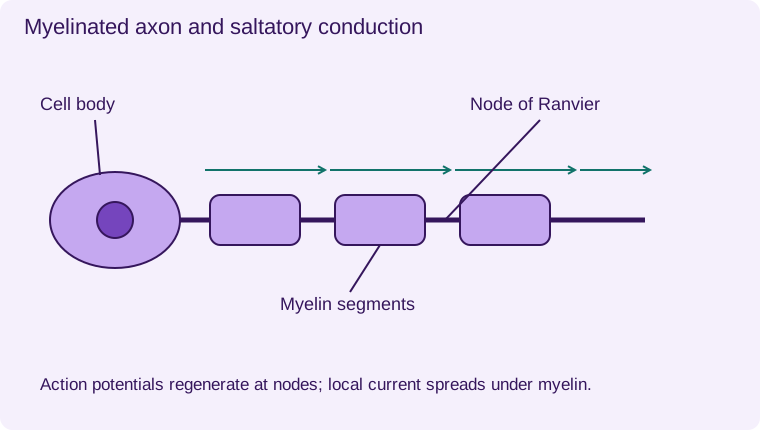
Myelin is a multilayered, lipid-rich sheath formed by glial cells. In the peripheral nervous system, Schwann cells form myelin segments. In the central nervous system, oligodendrocytes provide myelin. Gaps between adjacent segments are nodes of Ranvier, where voltage-gated channels are concentrated.
Myelin reduces current leakage and enables local depolarising current to spread rapidly beneath the sheath. Action potentials are regenerated at the nodes: saltatory conduction. The impulse does not physically leap through empty space. Demyelination can slow or block conduction by disrupting this arrangement.
Compare axons fairly
In non-myelinated axons, action potentials are regenerated continuously along the membrane. These axons may still be associated with supporting glial cells; non-myelinated does not mean unsupported. For axons of comparable diameter and conditions, myelination increases conduction speed. Diameter and temperature also influence speed, so control these when comparing experimental data.
A longer axon increases the distance a signal must travel, while speed determines how long it takes. Reaction time also includes receptor processing, synaptic delays and muscle activation, so it is not simply axon conduction time. Use the terms nerve, neurone and axon carefully: a peripheral nerve is a bundle of nerve fibres, not one cell.
- 1
- Dendrites
- 2
- Cell body and nucleus
- 3
- Axon
- 4
- Myelin segment
- 5
- Node of Ranvier: exposed axon
- 6
- Axon terminals
Action potentials regenerate at nodes; local current spreads under myelin. Schematic, not to scale.
Worked application and data skills
A 0.8 m pathway conducts at 40 m s⁻¹. Travel time = distance/speed = 0.8/40 = 0.020 s = 20 ms. At 2 m s⁻¹, the same distance takes 0.40 s. These are conduction times only; a complete behavioural response involves additional stages.
Common misconceptions
Myelin is not continuous across nodes. Non-myelinated axons still have membranes. A nerve contains many fibres.
Examination tips
Use a matched comparison: myelin, nodes, regeneration pattern and relative conduction speed.
Check your understanding
Five original questions. Use Tab to move between controls, arrow keys within a radio group, and Enter or Space on buttons. Feedback appears after Check Answer. Try Again clears the question. Attempts are not saved.
Sources and scope
Scope: Unit 1 A3.4.1. Original BTECRevision notes, schematics and questions. Independent resource; not endorsed by Pearson.
Official Pearson specification. Scientific review references are listed in the accompanying QA report.
EXAM RESOURCES
Biology exam resources
Apply your knowledge with written practice.
Browse exam resources