Gram-Positive and Gram-Negative Bacteria: Antibiotic Responses
FREE BIOLOGY REVISION · UNIT 1 · A1.4
Pearson BTEC Level 3 AAQ Applied Science · Issue 7, May 2026
Revision notes
Two cell-envelope patterns
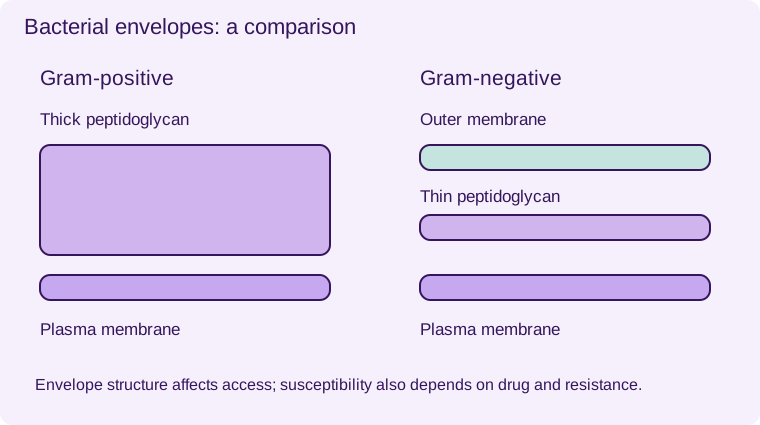
Gram-positive bacteria have a thick peptidoglycan wall outside their plasma membrane and no Gram-negative-type outer membrane. Gram-negative bacteria have a much thinner peptidoglycan layer in the periplasm between the plasma membrane and an outer membrane. The outer membrane contains lipopolysaccharide and porins, which control entry of some substances.
Gram staining reflects these envelope differences. Crystal violet and iodine form a complex; after decolourisation, Gram-positive cells generally retain purple staining. Gram-negative cells lose it and take up the counterstain, appearing pink or red. Staining is a classification aid, not a direct test of which antibiotic will work.
Link envelope structure to a drug target
Beta-lactam antibiotics such as penicillins inhibit bacterial enzymes involved in peptidoglycan cross-linking. In susceptible, growing bacteria this weakens the wall and can lead to osmotic lysis. Gram-negative outer membranes can restrict access of some drugs; particular antibiotics may pass through porins or use other routes. The effect depends on the exact antibiotic and organism.
Resistance can result from beta-lactamase enzymes, altered targets, reduced permeability or efflux pumps. Gram-positive bacteria can be resistant and Gram-negative bacteria can be susceptible. Do not convert a general envelope comparison into the false rule that all Gram-negative bacteria resist all antibiotics. Antibiotics acting on ribosomes have different targets from wall-active antibiotics. Viruses have no peptidoglycan or bacterial ribosomes.
Interpret a susceptibility investigation
An agar diffusion test places antibiotic discs on a standardised bacterial lawn. A clear inhibition zone indicates reduced visible growth under those conditions. Keep inoculum, medium, incubation conditions and disc content controlled. Diameter alone should not be used to rank different antibiotics without validated interpretive standards: diffusion rates and disc amounts differ.
Use fictional results in revision, not clinical recommendations. If the same drug at the same dose produces a larger zone with strain X than strain Y, the result supports greater inhibition of X in that assay, but a clinical susceptibility classification requires the appropriate method and breakpoints.
Gram-positive
- 1
- Thick peptidoglycan
- 2
- Plasma membrane
Gram-negative
- 1
- Outer membrane
- 2
- Thin peptidoglycan
- 3
- Plasma membrane
Envelope structure affects access; susceptibility also depends on the antibiotic and resistance mechanisms. Schematic, not to scale.
Worked application and data skills
Fictional strains X and Y receive identical discs of antibiotic P under matched conditions. Zones are 24 mm and 8 mm respectively. X shows greater inhibition in this test. You cannot conclude that all Gram-positive bacteria behave like X or that P will necessarily cure an infection in a person.
Common misconceptions
Gram status does not guarantee susceptibility. Antibiotics do not treat viruses through bacterial wall targets. A clear zone is not proof that every cell was killed.
Examination tips
Connect drug access, target and resistance. Separate what the results show from what they cannot establish.
Check your understanding
Five original questions. Use Tab to move between controls, arrow keys within a radio group, and Enter or Space on buttons. Feedback appears after Check Answer. Try Again clears the question. Attempts are not saved.
Sources and scope
Scope: Unit 1 A1.4. Original BTECRevision notes, schematics and questions. Independent resource; not endorsed by Pearson.
Official Pearson specification. Scientific review references are listed in the accompanying QA report.
EXAM RESOURCES
Biology exam resources
Apply your knowledge with written practice.
Browse exam resources