Protein Structure: From Peptide Bonds to Haemoglobin
FREE BIOLOGY REVISION · UNIT 1 · B3.1
Pearson BTEC Level 3 AAQ Applied Science · Issue 7, May 2026
Learning objectives
By the end of this topic you should be able to:
- Describe primary, secondary, tertiary and quaternary protein structure.
- Explain peptide-bond formation and interactions maintaining protein shape.
- Use haemoglobin to relate protein organisation to function.
Revision notes
Primary and secondary structure
Amino acids contain an amino group, a carboxyl group and a variable R group attached to a central carbon. Condensation between amino acids forms a peptide bond and releases water. A polypeptide has an amino-acid sequence: its primary structure. The sequence determines which interactions can occur during folding; a change in sequence can therefore alter function.
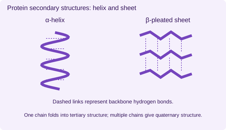
Secondary structure includes α-helices and β-pleated sheets, stabilised by hydrogen bonds between backbone groups. These are local regular arrangements. They are not created by disulfide bonds between every adjacent amino acid, and a single protein can contain both types.
Tertiary structure
Tertiary structure is the overall three-dimensional shape of one polypeptide. Hydrogen bonds, ionic interactions, disulfide bridges between cysteine side chains, van der Waals forces and hydrophobic interactions contribute. Disulfide bridges are covalent; most other listed interactions are individually weaker but numerous. Folding brings amino acids far apart in the sequence close together in space.
The distribution of R groups influences solubility and the arrangement of binding sites. Heating or extreme pH can disrupt interactions, changing shape and function: denaturation. The peptide backbone usually remains intact under ordinary denaturing conditions. Hydrolysis of peptide bonds is a different process.
Quaternary structure
Quaternary structure describes the association of more than one polypeptide subunit. Adult haemoglobin has four subunits, each associated with a haem group containing iron that binds oxygen. The haem groups are non-protein components. Not every protein has quaternary structure: a single functional polypeptide can have only primary, secondary and tertiary organisation.
Globular and fibrous
Globular proteins are generally compact and often soluble, suited to transport, catalysis and signalling. Fibrous proteins have elongated structures and provide mechanical roles; collagen is an example. These are broad structure–function categories, not a rule that every protein in blood must have the same shape or every structural protein must be chemically identical.
Enlarge diagram
Swipe the larger view, or focus it and use the arrow keys.
Amino-acid sequence joined by peptide bonds
α-helices and β-sheets with backbone hydrogen bonds
One polypeptide folds into an overall three-dimensional structure
Multiple polypeptides associate, as in haemoglobin
Worked application and data skills
A chain of 50 amino acids contains 49 peptide bonds and releases 49 water molecules during its initial assembly. If four separately synthesised polypeptides associate, that gives quaternary organisation; the subunit association is not automatically another long peptide chain. A mutation changing an R group can alter ionic interactions and folding even when chain length is unchanged.
Common misconceptions
Denaturation usually changes folding without hydrolysing the primary structure. Hydrogen bonds stabilise secondary structure. Haemoglobin contains four polypeptide subunits, not four amino acids.
Examination tips
Name the structural level and the relevant interaction. Distinguish peptide bonds from disulfide bridges.
Check your understanding
Five original questions. Use Tab to move between controls, arrow keys within a radio group, and Enter or Space on buttons. Feedback appears after Check Answer. Try Again clears the question. Attempts are not saved.
Sources and scope
Scope: Unit 1 B3.1. Original BTECRevision notes, schematics and questions. Independent resource; not endorsed by Pearson.
Official Pearson specification. Scientific review references are listed in the accompanying QA report.
Take Your Learning Further
Practise
Build Your Confidence
Reinforce your understanding with our topic-based workbook. Every workbook page links back to its matching online lesson, so you can revisit explanations whenever you need support.
Explore Workbook – BiologyMaster
Get Exam Ready
Apply your knowledge with exam-style practice papers and detailed mark schemes. Identify what you’ve mastered and what to revise next.
Practice Papers – BiologyEXAM RESOURCES
Biology exam resources
Apply your knowledge with written practice.
Browse exam resources