Skeletal Muscle Fibres and Sarcomeres
FREE BIOLOGY REVISION · UNIT 1 · A3.3.1
Pearson BTEC Level 3 AAQ Applied Science · Issue 7, May 2026
Revision notes
From muscle to filament
A skeletal muscle contains bundles of long muscle fibres. Each fibre is a multinucleated cell bounded by the sarcolemma. Its cytoplasm, or sarcoplasm, contains mitochondria and many myofibrils. Myofibrils consist of repeating sarcomeres, the contractile units between adjacent Z lines. Aligned sarcomeres give skeletal muscle its striated appearance.
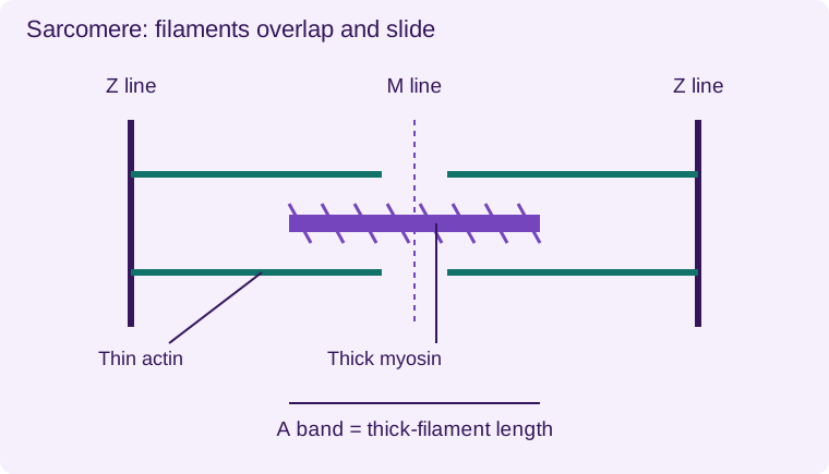
Thin filaments contain actin and associated regulatory proteins. Thick filaments contain myosin with projecting heads. Thin filaments anchor to Z lines; thick filaments occupy the central region around the M line. The A band spans the length of thick filaments; the I band contains thin filaments without thick-filament overlap. The H zone is the central thick-filament-only region.
Sliding-filament mechanism
An electrical signal travels along the sarcolemma and into T tubules, leading to calcium release from the sarcoplasmic reticulum. Calcium binds to troponin, shifting tropomyosin so that myosin-binding sites on actin become accessible. Myosin heads bind actin and generate force through repeated cross-bridge cycles.
ATP binding allows a myosin head to detach. ATP hydrolysis energises it for another cycle. Calcium is actively returned to the sarcoplasmic reticulum during relaxation. ATP is therefore needed for both cycling and restoration, not merely for the power stroke in a vague sense.
What shortens?
Thin and thick filaments slide past one another; the filaments do not themselves become shorter. Z lines move closer, the I band and H zone narrow, and the A band stays approximately constant. A whole muscle shortens because many sarcomeres shorten together. A cell diagram should distinguish a complete fibre, a myofibril and an individual sarcomere.
Connect structure to use: numerous mitochondria support ATP supply, the sarcoplasmic reticulum permits rapid calcium control, and organised parallel filaments transmit force. Detailed motor-unit recruitment is useful background but does not replace the required microscopic structure.
- 1
- Z line
- 2
- Z line
- 3
- M line
- 4
- Thin actin filament
- 5
- Thick myosin filament
A band = thick-filament length. Filaments overlap and slide; the filaments themselves do not shorten. Schematic, not to scale.
Worked application and data skills
A sarcomere shortens from 2.4 µm to 1.8 µm. Percentage shortening = (2.4 − 1.8)/2.4 × 100 = 25%. The myosin filament length remains constant; increased overlap accounts for the change. An image showing a narrower I band with an unchanged A band supports contraction.
Common misconceptions
Filaments slide rather than shrink. A muscle fibre is one long cell. ATP is required to detach myosin and restore calcium gradients.
Examination tips
For a micrograph, identify Z lines, A band and I band before interpreting contraction.
Check your understanding
Five original questions. Use Tab to move between controls, arrow keys within a radio group, and Enter or Space on buttons. Feedback appears after Check Answer. Try Again clears the question. Attempts are not saved.
Sources and scope
Scope: Unit 1 A3.3.1. Original BTECRevision notes, schematics and questions. Independent resource; not endorsed by Pearson.
Official Pearson specification. Scientific review references are listed in the accompanying QA report.
EXAM RESOURCES
Biology exam resources
Apply your knowledge with written practice.
Browse exam resources