Recognising Cells and Organelles in Micrographs
FREE BIOLOGY REVISION · UNIT 1 · A1.2
Pearson BTEC Level 3 AAQ Applied Science · Issue 7, May 2026
Revision notes
Read an image systematically
A photomicrograph is an image obtained using light microscopy. An electron micrograph uses electrons and can resolve much finer detail. Transmission electron microscopy (TEM) shows internal sections; scanning electron microscopy (SEM) gives surface detail. Most biological electron micrographs are originally greyscale; colours may have been added later. Colour is therefore a poor identification rule.
Magnification describes image size relative to actual size. Resolution is the minimum separation at which two points can be distinguished. Enlarging a blurred image increases magnification without revealing more detail. Light microscopy can show whole cells, nuclei, chloroplasts and some larger structures, but does not normally resolve individual ribosomes or membrane bilayers.
Use visible diagnostic features
Identify a nucleus by its envelope and chromatin; a dark nucleolus may be visible within it. Mitochondria show an outer boundary and folded cristae. Chloroplasts show internal thylakoids, often in stacked grana, and may contain starch grains. RER appears as flattened membranes with attached ribosomes; smooth ER lacks these attached particles. Golgi stacks are curved flattened sacs with nearby vesicles. A large pale compartment bounded by a tonoplast can be a vacuole.
Cell walls form relatively thick, regular boundaries in many plant sections. A membrane alone is much thinner. Cilia may appear as surface projections or, in cross-section, as microtubule arrangements. Small dark spots may be ribosomes, but do not identify every dark dot as a ribosome without scale and context.
Why a real image differs from a diagram
Micrographs are sections through three-dimensional structures. The same organelle can appear round, elongated or incomplete according to the cutting plane. Fixation, dehydration and staining can distort structures. A single section may miss the nucleus entirely. Absence from one image is not proof that the living cell lacks that structure.
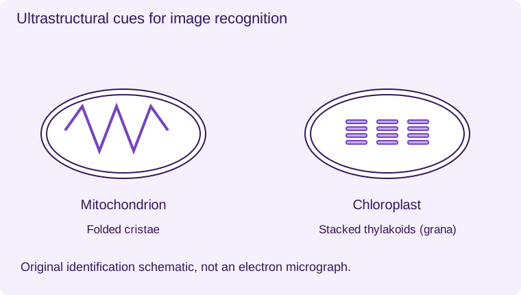
The original schematic identification plate provided here reproduces structural cues, not a real specimen or a Pearson image. The genuine TEM and light micrograph below provide additional practice. The schematic is not a real specimen. Compare the real images with the structural cues rather than relying on colour.
Mitochondrion
Double boundary and folded cristae
Chloroplast
Double boundary and stacked thylakoids (grana)
Original identification schematics, not electron micrographs. Actual micrographs in the lesson are unchanged.
Nucleus
- 1
- Nucleolus
- 2
- Double nuclear envelope with pores
Rough ER
- 1
- Membrane cisternae
- 2
- Attached 80S ribosomes
Golgi and vesicles
- 1
- Golgi stacks
- 2
- Transport vesicle
- 3
- Lysosome: digestive enzymes
Smooth ER lacks attached ribosomes. Membrane-bound vesicles transport material between compartments.
Worked application and data skills
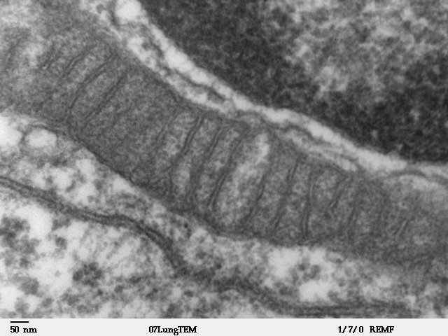
Image X contains an oval organelle with a double envelope and numerous internal folds. Identify it as a mitochondrion and cite the cristae. If instead it contains organised stacks of internal membranes, a chloroplast is more likely. Check the scale: an isolated small dark particle does not have enough structure to justify either identification.
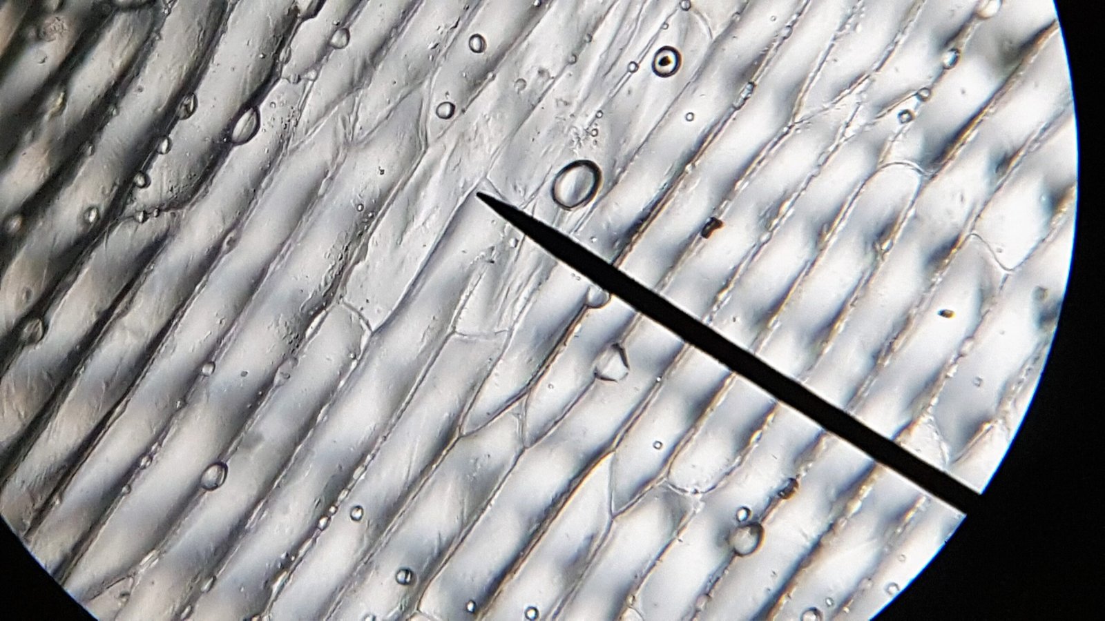
Real-image task 1: inspect the TEM. Identify the large oval organelle using its internal cristae, then distinguish the surrounding cytoplasm. Real-image task 2: inspect the onion photomicrograph. Trace the repeated cell-wall boundaries. Do not infer that nuclei or chloroplasts must be visible in this image. The TEM includes an original scale bar; use it only when its label and endpoints can be read reliably. The onion image does not provide a calibrated scale bar for size calculations. Numerical practice uses the separate original scale-bar diagram.
Common misconceptions
Magnification and resolution are not interchangeable. Electron micrographs are not naturally purple. A section can miss an organelle.
Examination tips
For an identification plus reason, name the organelle and cite a feature actually visible in the image.
Check your understanding
Five original questions. Use Tab to move between controls, arrow keys within a radio group, and Enter or Space on buttons. Feedback appears after Check Answer. Try Again clears the question. Attempts are not saved.
Sources and scope
Scope: Unit 1 A1.2. Original BTECRevision notes, schematics and questions. Independent resource; not endorsed by Pearson.
Official Pearson specification. Scientific review references are listed in the accompanying QA report.
EXAM RESOURCES
Biology exam resources
Apply your knowledge with written practice.
Browse exam resources