Peripheral Sensory Pathways and Autonomic Feedback
FREE BIOLOGY REVISION · UNIT 1 · C3.4.4
Pearson BTEC Level 3 AAQ Applied Science · Issue 7, May 2026
Revision notes
Central and peripheral organisation
The central nervous system comprises brain and spinal cord. The peripheral nervous system includes nerves and ganglia outside them. Peripheral sensory, or afferent, pathways carry information towards the CNS. Motor, or efferent, pathways carry output from the CNS towards effectors. Autonomic motor pathways regulate smooth muscle, cardiac muscle and glands.
Visceral sensory fibres report conditions within the body, including pressure and chemical changes. These afferent pathways work with autonomic control but should not be confused with outgoing sympathetic or parasympathetic motor fibres. The specification’s reference to relaying information to the brain is best understood by distinguishing incoming sensory information from outgoing autonomic responses.
A reflex loop
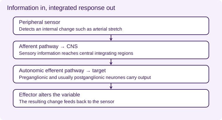
In the arterial-pressure example, stretch-sensitive baroreceptors detect a pressure change. Sensory signals travel through peripheral cranial-nerve pathways to the brainstem. Cardiovascular centres integrate the information and alter autonomic output to the heart and vessels. The resulting change in cardiac output and resistance opposes the initial pressure deviation.
Some reflex integration can occur in the spinal cord or peripheral enteric networks, while higher centres can modify responses. Not every reflex must involve conscious awareness. Conversely, saying that the brain is involved does not mean the person chooses the response voluntarily.
Two-neurone autonomic output
Many autonomic motor routes use a preganglionic neurone from the CNS and a postganglionic neurone from an autonomic ganglion to the target. This differs from the usual single somatic motor neurone reaching skeletal muscle from the CNS. The adrenal medulla is a specialised exception receiving preganglionic sympathetic input directly.
Interpret a pathway lesion
Damage to sensory input can prevent accurate detection of a disturbance even if the effector is intact. Damage to motor output can prevent a response despite normal sensing. Failure can therefore occur at multiple positions. To localise it, compare detection, central processing and effector responses rather than assuming every abnormal response reflects a receptor problem.
Detects an internal change such as arterial stretch
Sensory information reaches central integrating regions
Preganglionic and usually postganglionic neurones carry output
The resulting change feeds back to the sensor
Worked application and data skills
A model raises arterial pressure and records increased sensory firing but no expected heart-rate change. This suggests sensing occurred, while integration, motor output or cardiac response may be impaired. It does not locate the fault uniquely. Directly testing the effector and recording autonomic output would narrow the possibilities.
Common misconceptions
Afferent means towards the CNS; efferent means away. Autonomic motor output is not the same as visceral sensory input. Automatic responses need not be conscious.
Examination tips
Trace information into the CNS before tracing the response out. Identify the direction of each pathway.
Check your understanding
Five original questions. Use Tab to move between controls, arrow keys within a radio group, and Enter or Space on buttons. Feedback appears after Check Answer. Try Again clears the question. Attempts are not saved.
Sources and scope
Scope: Unit 1 C3.4.4. Original BTECRevision notes, schematics and questions. Independent resource; not endorsed by Pearson.
Official Pearson specification. Scientific review references are listed in the accompanying QA report.
Take Your Learning Further
Practise
Build Your Confidence
Reinforce what you’ve learned with our topic-based workbook. Every workbook page links back to its matching online lesson, so help is always at hand.
Explore Workbooks – BiologyMaster
Get Exam Ready
Challenge yourself with exam-style practice papers and detailed mark schemes. Apply your knowledge, identify areas to improve and build exam confidence.
Practice Papers – BiologyEXAM RESOURCES
Biology exam resources
Apply your knowledge with written practice.
Browse exam resources